Magnetic Bead Separation: Principles and Streptavidin Bead Applications
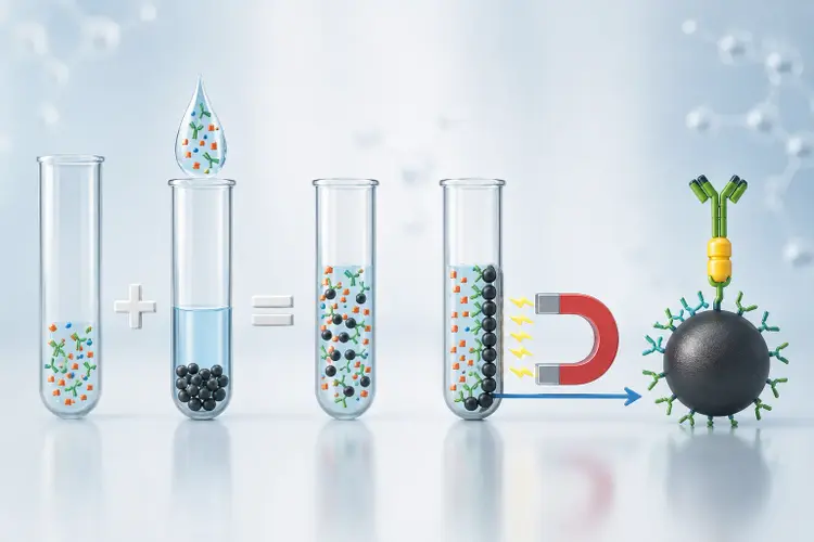
Separation using magnetic beads is a widely used approach for isolating defined targets from complex biological samples. The same core principle also supports streptavidin magnetic beads, which are used to capture biotinylated proteins and peptides for purification and downstream analysis. Researchers across immunology, cancer biology, stem cell research, and translational science use magnetic separation because it is fast, gentle, scalable, and compatible with workflows that need clean enrichment before downstream analysis (1). When the method is designed carefully, it can support strong target selectivity in addition to fitting smoothly into tube, plate, column, and automated sample preparation formats (1, 4, 7).
The value of magnetic separation starts with its simplicity. A magnetic particle is linked to a target through an antibody, ligand, or affinity partner, after which a magnetic field is applied so the bound fraction can be retained while unwanted material is removed (1). That broad principle is important in cell isolation, and it is also the foundation for one of the most useful affinity workflows in modern sample preparation: streptavidin magnetic beads for the isolation, cleanup, and purification of proteins and peptides through biotin-based capture (3–5).
What Is Magnetic Bead-Based Cell Separation?
In positive selection, the intended cell population is directly labeled and retained. In negative, or untouched, selection, unwanted populations are labeled and removed so the desired cells remain untagged in the final fraction (1).
Labs typically judge performance using purity, recovery, and viability. These three measures capture how selective, complete, and gentle a workflow really is, irrespective of whether the target is a cell population or a biotinylated biomolecule (1).
Principle and Working Mechanism of Magnetic Bead-Based Cell Separation
The working mechanism behind cell separation using magnetic beads depends on selective binding first, then controlled retention in a magnetic field. The bead surface is coated with an antibody, ligand, or affinity chemistry that recognizes the target of interest. After incubation, the sample is placed in a magnetic field so the labeled fraction is held in place while unbound material is removed through aspiration, washing, or transfer steps (1, 4). This makes the process highly adaptable across cell biology, immunocapture, and sample preparation workflows.
An important physical feature of many magnetic particles is superparamagnetism. Superparamagnetic particles respond strongly to an applied magnetic field but do not retain residual magnetization once the field is removed, which helps minimize bead-to-bead attraction after separation, reduce aggregation, and improve resuspension consistency between process steps (1). In column-based formats, a ferromagnetic matrix can intensify the local magnetic field and improve retention of small, labeled targets, especially when very fine particles are used (1).
Once that general mechanism is understood, streptavidin magnetic beads become easier to place in context. Streptavidin-coated magnetic beads rely on a very tight biotin-binding pair that is widely used for selective capture in life science workflows (3). Their strong binding behavior and dependable performance make them well-suited for isolating biotinylated proteins and peptides before cleanup, enrichment, and downstream analysis (3, 5).
Method performance still depends on practical variables. Antigen or ligand density, bead-to-target ratio, sample concentration, viscosity, binding kinetics, incubation time, washing stringency, and sample load all affect the final result, so fit-for-purpose testing matters even when the core chemistry is well established. Incubation conditions should also be optimized for the specific assay, since the best temperature and timing can vary by target and workflow (1, 4, 5).
Key Benefits of Using Magnetic Beads for Separation
Magnetic beads for cell separation are attractive because they combine target specificity with operational flexibility. For researchers working with proteins and peptides, streptavidin magnetic beads extend those same benefits into affinity purification and analytical sample preparation workflows (3–5).
- They support selective enrichment of target cell populations from complex biological matrices (1).
- They can be integrated into gentle workflows that preserve cell quality when viability matters (1).
- They fit manual, semi-automated, and automated sample preparation formats, which helps labs scale from method development to higher-throughput processing (4, 6–8).
- They work well with downstream tools such as flow cytometry, culture, liquid chromatography-tandem mass spectrometry (LC-MS/MS), and other molecular assays that depend on cleaner inputs (4, 5, 6, 8).
- In streptavidin-based workflows, selective capture of biotinylated targets and thorough wash steps reduce background and prepare cleaner samples for LC-MS/MS and related analytical work (3–5).
- They are useful for enrichment of low-abundance analytes, where washing quality and selective capture can materially improve the analytical value of the final fraction (4, 7, 8).
Step-by-Step Process of Magnetic Bead-Based Separation
A typical workflow of cell separation via magnetic beads begins with sample preparation and ends with a retained or released target fraction ready for analysis. The exact details depend on whether the goal is intact cell isolation or affinity capture of biomolecules, but the broad process is similar across both use cases (1, 4).
1. Preparation of the Sample
The sample should be prepared in a form that supports target access and controlled binding. For cell workflows, that usually means a clean single-cell suspension with minimal clumping, as in applications such as T-cell enrichment, PBMC subset isolation, stem cell enrichment, and rare-cell capture. For protein and peptide workflows, it means a buffer system and sample condition compatible with the capture chemistry and the downstream assay (1, 4, 6). Overloaded or poorly clarified samples can make capture less selective and washing less effective, so basic sample quality still matters before any magnetic step begins (1, 5).
2. Binding Cells to Magnetic Beads
In classical cell separation, most commercial magnetic beads are pre-functionalized with an antibody or ligand that recognizes a marker on the target cell. Positive selection retains the labeled target cells, while untouched workflows remove other populations and leave the target unlabeled (1).
3. Affinity Capture with Streptavidin Magnetic Beads
Beyond cell separation, this is where the workflow shifts into affinity purification for proteins and peptides. Streptavidin magnetic beads are used to pull down biotinylated proteins and peptides through a highly selective binding system that is well-suited to affinity capture workflows (3, 5). Phenomenex’s streptavidin-coated Biozen MagBeads are positioned for protein and peptide isolation, clean-up, and purification. The magnetic beads procedure describes magnetic beads as a rapid way to isolate and purify proteins and peptides through affinity binding.
4. Magnetic Separation
After binding, the sample is placed in a magnetic separator so the labeled fraction is retained and the rest of the material can be removed. Wash-stringency optimization is a key control point because it helps balance purity against recovery, and overly aggressive washes can reduce yield. This step is simple in principle, but wash quality is crucial. In proteomics and peptide enrichment workflows, wash steps help remove nonspecific material and reduce sample complexity before downstream analysis (4, 7, 8).
5. Elution of Target Cells or Biomolecules
The final step depends on the assay design. , and target release is not universally available across magnetic bead platforms. Some workflows keep the target bound to the beads for a downstream analysis, while others allow elution or bead removal only in platforms built for that purpose (2, 4, 6). For cell workflows, quality control often includes flow cytometry with a viability dye so purity, recovery, and viability can be measured together rather than inferred from yield alone (1).
Applications of Magnetic Bead-Based Separation
Once general magnetic isolation is understood, the most commercially and scientifically relevant shift now is toward streptavidin magnetic beads for affinity-based capture. These workflows use the same core magnetic principle and apply it to the selective isolation of biotinylated proteins, peptides, antibodies, and related biomolecules before downstream analytical work (3–5).
Protein and Peptide Purification
Streptavidin magnetic beads are widely used for affinity capture of biotinylated proteins, peptides, and antibodies. They support immunocapture workflows, cleanup before LC-MS/MS, and any workflow where selective isolation of a biotinylated target from a complex matrix is the goal (3, 5). Because the binding interaction is stable and selective, researchers can use wash steps to remove background effectively before the sample reaches the analytical step.
Immunoprecipitation and Pull-Down Assays
Magnetic beads are widely used in immunoprecipitation, co-immunoprecipitation, and pull-down assays for studying protein-protein interactions and related complexes. Streptavidin bead pull-down methods can capture biotin-tagged interaction partners in a selective format, helping researchers isolate bound material for follow-up characterization (2, 5). This makes magnetic beads useful not only for preparative isolation but also for mechanistic and biomarker-focused workflows.
Sample Preparation for Proteomics and Molecular Biology
Magnetic bead workflows also support sample preparation for proteomics and molecular biology by combining enrichment, washing, and downstream compatibility in a format that can be manual or automated. Paramagnetic bead-based workflows, including single-tube and single-pot strategies, are used to reduce sample loss, improve throughput, and support LC-MS/MS analysis from limited or complex samples (4, 6–8).
Biomolecule Enrichment and Cleanup
Magnetic bead-based enrichment is especially helpful for low-abundance targets that would otherwise be difficult to recover cleanly from complex matrices. Selective capture, thorough washing, and compatibility with downstream digestion or elution steps can improve assay cleanliness and analytical confidence without requiring a complicated workflow (4, 5, 7, 8). These workflows can be run in manual tube-based formats or adapted to higher-throughput platforms, which broadens their usefulness across research and sample preparation settings (4, 7, 8).
Workflow Considerations
The clearest checkpoints for a magnetic bead workflow are purity, recovery, and background level at the end of each run. For cell workflows, flow cytometry with a viability dye gives a direct readout of all three. For protein and peptide workflows, analytical confirmation through LC-MS/MS or a plate-based assay is the most reliable way to verify that the enrichment step worked as expected (1, 4, 6–8).
FAQs on Magnetic Bead-Based Separation
What types of cells can be separated using magnetic beads?
Many cell types can be separated if a suitable marker or depletion strategy is available, including immune cells, stem and progenitor populations, and disease-relevant subpopulations from mixed biological samples.
How do I choose the right magnetic beads for my experiment?
The choice depends on the target, the capture chemistry, and the downstream assay. Researchers should consider whether the workflow needs positive or untouched selection, direct affinity capture, a given binding capacity, or compatibility with later steps such as flow cytometry or LC-MS/MS.
How to separate magnetic beads from cells?
That depends on the specific system. Some workflows keep the beads attached, while others support release or post-capture removal. Bead removal should only be described when the chemistry and workflow explicitly support it.
How do I choose between positive and negative selection?
Positive selection enriches the target population directly, whereas negative selection removes unwanted cells and leaves the desired cells untouched. The better choice depends on whether the downstream assay requires maximum purity, minimal bead exposure, preserved native function, or the least possible manipulation of the target cells.
What equipment is used in magnetic separation?
Common tools include magnetic racks, magnetic stands, separator columns, wash vessels, filtration accessories, and downstream QC tools such as flow cytometers. Automated platforms may also be used for higher-throughput enrichment and sample preparation workflows.
What are the advantages of separation using magnetic beads?
The main advantages are selectivity, speed, gentle handling, scalability, and compatibility with downstream analysis. The same strengths also support affinity workflows for proteins and peptides when the bead chemistry matches the assay design.
How do streptavidin magnetic beads help with protein purification?
Streptavidin magnetic beads capture biotinylated proteins and peptides through a very stable, selective binding interaction. That makes them useful for affinity isolation, enrichment, and cleanup before LC-MS/MS or related analytical steps.
References
- Plouffe, B. D., Murthy, S. K., & Lewis, L. H. (2015). Fundamentals and application of magnetic particles in cell isolation and enrichment. Reports on Progress in Physics, 78(1), 016601. https://pmc.ncbi.nlm.nih.gov/articles/PMC4310825/
- Xu, X., Dong, C., Hoffmann, T., & Goh, P. Y. (2017). Streptavidin bead pulldown assay to determine protein-protein interactions. Bio-Protocol, 7(22), e2613. https://pmc.ncbi.nlm.nih.gov/articles/PMC5790316/
- Chivers, C. E., Koner, A. L., Lowe, E. D., & Howarth, M. (2011). How the biotin-streptavidin interaction was made even stronger. Biochemical Journal, 435(1), 55–63. https://pmc.ncbi.nlm.nih.gov/articles/PMC3062853/
- Becker, A., Wiest, M., Telek, A., et al. (2022). Transforming chemical proteomics enrichment into a high-throughput method using optimization of a single magnetic bead handling step. JACS Au, 2(8), 1862–1877. https://pmc.ncbi.nlm.nih.gov/articles/PMC9326820/
- Berg Luecke, L., & Gundry, R. L. (2021). Assessment of streptavidin bead binding capacity to improve quality of streptavidin-based enrichment studies. Journal of Proteome Research, 20(7), 3155–3168. https://pmc.ncbi.nlm.nih.gov/articles/PMC8116117/
- Hughes, C. S., Foehr, S., Garfield, D. A., Furlong, E. E. M., Steinmetz, L. M., & Krijgsveld, J. (2014). Ultrasensitive proteome analysis using paramagnetic bead technology. Molecular Systems Biology, 10, 757. https://pmc.ncbi.nlm.nih.gov/articles/PMC4299378/
- Kanshin, E., & Thibault, P. (2015). Efficient sample processing for proteomics applications: Are magnetic beads the answer? Molecular Systems Biology, 10, 757 commentary. https://pmc.ncbi.nlm.nih.gov/articles/PMC4299379/
- Anderson, N. L., Jackson, A., Smith, D., Hardie, D., Borchers, C. H., & Pearson, T. W. (2009). SISCAPA peptide enrichment on magnetic beads using an in-line bead trap device. Molecular & Cellular Proteomics, 8(5), 995–1005. https://pubmed.ncbi.nlm.nih.gov/19196707/